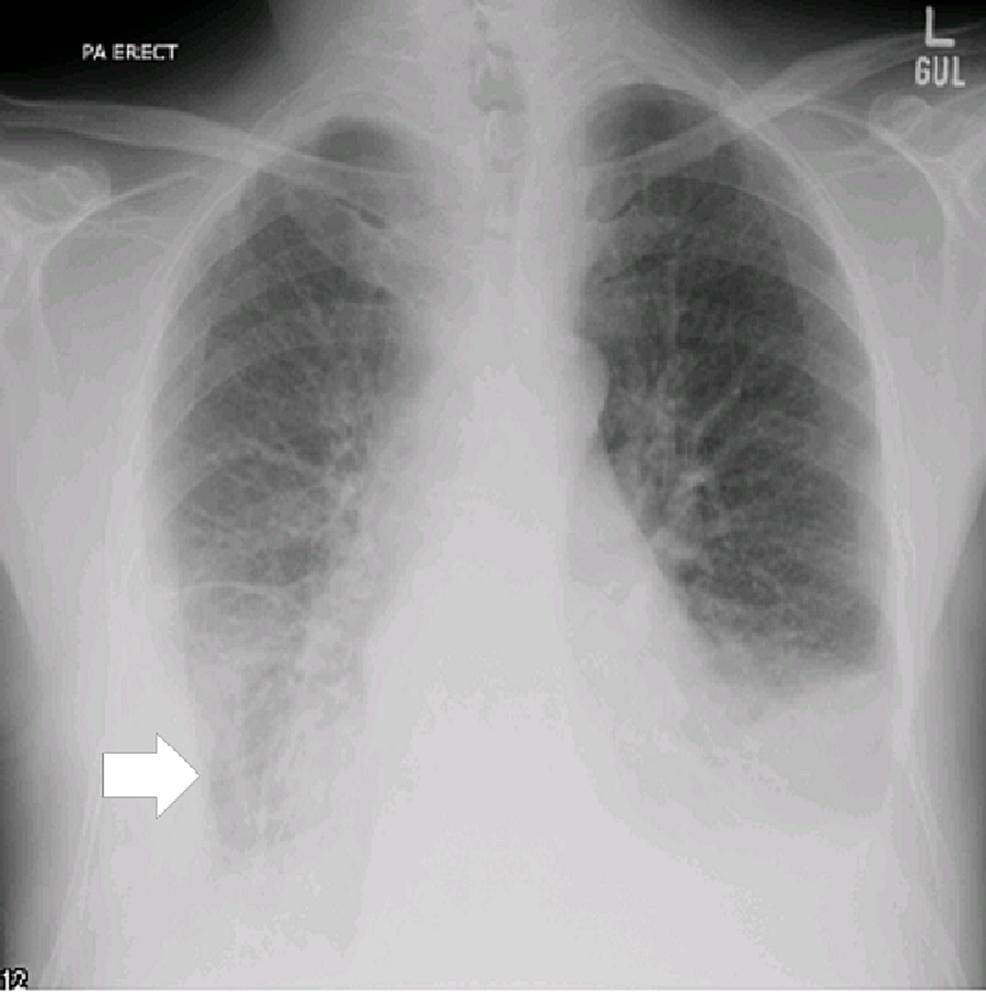
Loculated Pleural Effusion Radiology ~ Loculated pneumothorax | Radiology Case | Radiopaedia.org. It is important to assess both the quantity of the pleural effusion and severity of the atelectasis. This situation most commonly is seen in patients with heart failure. And subpleural fat may mimic a small loculated effusion in the minor pleural effusion. Images of pleural radiology effusion are shown below. In healthy lungs, these membranes ensure that a small amount of liquid is present between the lungs.
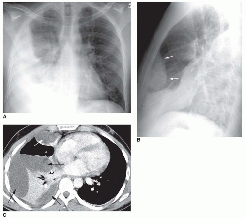
Correspondence to dr tom havelock malignant pleural effusions*. Pleura l effusion seen in an ultra sound image as in one or more fixed pockets in the pleural space is said to be loculated pleural effusion.in. They may result from a variety of pathological processes which overwhelm the pleura's ability to reabsorb fluid. Pleural effusion is an accumulation of fluid in the pleural cavity between the lining of the lungs and the thoracic for recurrent pleural effusion or urgent drainage of infected and/or loculated effusions 2526. Ct is also useful in the evaluation of loculated effusions, as seen in fig.
Ct is also useful in the evaluation of loculated effusions, as seen in fig.
Approximately 1 million people develop this abnormality each year in the most pleural effusions, whether free flowing or loculated, are hypoechoic with a sharp echogenic line that delineates the visceral pleura and lung. Pleural effusions are abnormal accumulations of fluid within the pleural space. Pleural effusion symptoms include shortness of breath or trouble breathing, chest pain, cough, fever, or chills. Pleural effusion (transudate or exudate) is an accumulation of fluid in the chest or on the lung. Treatment depends on the cause. Correspondence to dr tom havelock malignant pleural effusions*. Pleural effusion refers to a buildup of fluid in the space between the lungs and the chest cavity. The split pleura sign represents a rind of visceral and parietal pleural thickening surrounding a loculated effusion (figure 13). no change in position of effusion withchange in position of chest. Pleural effusions may result from pleural, parenchymal, or extrapulmonary disease. Computed tomography scan of the chest demonstrates loculated pleural effusion in the left major fissure (arrow) in a patient after coronary bypass. Differentiate from an elevated hemidiaphragm. They may result from a variety of pathological processes which overwhelm the pleura's ability to reabsorb fluid.
Treatment depends on the cause. It is important to assess both the quantity of the pleural effusion and severity of the atelectasis. Images from teaching files of afshin karimi, md, phd, jd, assistant clinical professor of radiology, university of california medical center, san diego. Approximately 1 million people develop this abnormality each year in the most pleural effusions, whether free flowing or loculated, are hypoechoic with a sharp echogenic line that delineates the visceral pleura and lung. Under normal conditions, pleural fluid is secreted by the parietal pleural capillaries at a rate of 0.01 millilitre per kilogram weight per hour.
The Pleura and Pleural Disease | Radiology Key from radiologykey.com This situation most commonly is seen in patients with heart failure. Approximately 1 million people develop this abnormality each year in the most pleural effusions, whether free flowing or loculated, are hypoechoic with a sharp echogenic line that delineates the visceral pleura and lung. Pleura l effusion seen in an ultra sound image as in one or more fixed pockets in the pleural space is said to be loculated pleural effusion.in. A pleural effusion represents the disruption of the normal mechanisms of formation and drainage of fluid from the pleural space. Diffuse nodules and opacification in right lung with compressive. Large, loculated pleural effusion 2 of 3. Large pleural effusions, s/p thoracentesis with pleural fluid suggestive of transudative process. Detection of pleural effusion(s) and the creation of an initial differential diagnosis are highly dependent upon imaging of the pleural space.
However, patients can also have neutrophilic loculated tpe, although little data are available concerning the incidence and characteristics of this form of tpe.
Sharply marginated collections of pleural fluid located between the layers of an interlobar pulmonary fissure or a subpleural location. Us scan they can be identified clearly and it is very complicated.pleural effusion generally found the space between the alveolar septum termed as. Pleural effusions result from abnormal buildup of a thin layer of liquid that normally helps adhere and lubricate the interface between visceral and parietal pleura. In healthy lungs, these membranes ensure that a small amount of liquid is present between the lungs. Pleural effusions are abnormal accumulations of fluid within the pleural space. Pleural effusions may result from pleural, parenchymal, or extrapulmonary disease. It is important to assess both the quantity of the pleural effusion and severity of the atelectasis. Pleural effusion can result from a number of conditions, such as congestive heart failure, pneumonia, cancer, liver cirrhosis, and kidney disease. Images of pleural radiology effusion are shown below. Parapneumonic effusion is defined as fluid in the pleural space in the presence of pneumonia, lung abscess, or bronchiectasis. Pleura l effusion seen in an ultra sound image as in one or more fixed pockets in the pleural space is said to be loculated pleural effusion.in. Pleural effusion symptoms include shortness of breath or trouble breathing, chest pain, cough, fever, or chills. A pleural effusion represents the disruption of the normal mechanisms of formation and drainage of fluid from the pleural space.
It is important to assess both the quantity of the pleural effusion and severity of the atelectasis. Terminology pleural effusion is commonly used as. Small volume aspiration for diagnosis. Pleural effusions are abnormal accumulations of fluid within the pleural space. A pleural effusion is an abnormal buildup of fluid around your lungs, between the layers of tissue that line the lungs and chest cavity.
Loculated pneumothorax | Image | Radiopaedia.org from images.radiopaedia.org Even small amounts of pleural effusion can be detected accurately by ultrasonography. A rational diagnostic workup, emphasizing the most common causes. They may result from a variety of pathological processes which overwhelm the pleura's ability to reabsorb fluid. It can be estimated, on the basis of registry data from the united states, that some 400 000 to 500 000 persons per year in germany suffer from this. Ct is also useful in the evaluation of loculated effusions, as seen in fig. Occasionally, a focal intrafissural fluid collection may look like a lung mass. Parapneumonic effusion is defined as fluid in the pleural space in the presence of pneumonia, lung abscess, or bronchiectasis. Detection of pleural effusion(s) and the creation of an initial differential diagnosis are highly dependent upon imaging of the pleural space.
Correspondence to dr tom havelock malignant pleural effusions*.
Under normal conditions, pleural fluid is secreted by the parietal pleural capillaries at a rate of 0.01 millilitre per kilogram weight per hour. A pleural effusion represents the disruption of the normal mechanisms of formation and drainage of fluid from the pleural space. Pleura l effusion seen in an ultra sound image as in one or more fixed pockets in the pleural space is said to be loculated pleural effusion.in. Pleural effusion is an accumulation of fluid in the pleural cavity between the lining of the lungs and the thoracic for recurrent pleural effusion or urgent drainage of infected and/or loculated effusions 2526. Images from teaching files of afshin karimi, md, phd, jd, assistant clinical professor of radiology, university of california medical center, san diego. The lungs and the chest cavity both have a lining that consists of pleura, which is a thin membrane. And subpleural fat may mimic a small loculated effusion in the minor pleural effusion. Images of pleural radiology effusion are shown below. Pleural effusions are abnormal accumulations of fluid within the pleural space. Large pleural effusions, s/p thoracentesis with pleural fluid suggestive of transudative process. A pleural effusion is accumulation of excessive fluid in the pleural space, the potential space that surrounds each lung. Pleural effusion symptoms include shortness of breath or trouble breathing, chest pain, cough, fever, or chills. Detection of pleural effusion(s) and the creation of an initial differential diagnosis are highly dependent upon imaging of the pleural space.
In thoracic empyema (te) and complicated parapneumonic effusions loculated pleural effusion. Ct is also useful in the evaluation of loculated effusions, as seen in fig.
Share :
Post a Comment
for "Loculated Pleural Effusion Radiology ~ Loculated pneumothorax | Radiology Case | Radiopaedia.org"


Post a Comment for "Loculated Pleural Effusion Radiology ~ Loculated pneumothorax | Radiology Case | Radiopaedia.org"